Juvenile Arthritis & Bone Health Risk Calculator
Bone Health Risk Assessment
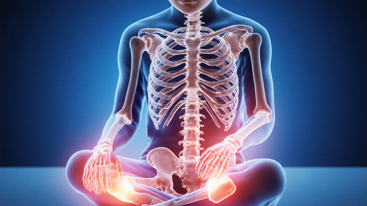
When a child is diagnosed with juvenile arthritis is a chronic inflammatory disorder that attacks the joints of children and teens, the focus often lands on pain relief and joint mobility. What many parents overlook is how the disease silently reshapes the developing skeleton, influencing bone density, growth plates, and long‑term skeletal strength. This article breaks down the link between the condition and bone health, explains why the connection matters, and offers concrete steps to keep those tiny bones as strong as possible.
What Is Juvenile Arthritis?
Juvenile arthritis (JA) is an umbrella term covering several types of arthritis that begin before age 16. The most common forms are:
- Oligoarticular JA - affects four or fewer joints, often the knees.
- Polyarticular JA - involves five or more joints, similar to adult rheumatoid arthritis.
- Systemic JA - combines joint inflammation with fever and rash.
All types share a hallmark: persistent inflammation of the synovial membrane, which produces extra fluid, swelling, and pain. The disease can flare up, subside, or linger for years, and the inflammatory process can directly interfere with bone remodeling-a natural balance of growth and breakdown that keeps the skeleton sturdy.
How Inflammation Impacts Growing Bones
Bone isn’t static; it’s constantly being rebuilt through two opposing cells: osteoclasts (break down bone) and osteoblasts (build bone). In a healthy child, this cycle aligns with growth spurts, ensuring the bones lengthen while staying dense.
Inflammation throws a wrench into that equilibrium. Pro‑inflammatory cytokines such as IL‑1, IL‑6, and TNF‑α accelerate osteoclast activity, causing more bone resorption. At the same time, they suppress osteoblasts, slowing new bone formation. The net effect is a lower bone mineral density (BMD) and, over time, a higher risk for fractures.
Common Bone‑Related Complications in Juvenile Arthritis
Because the skeleton is still maturing, the consequences of chronic inflammation show up in specific ways:
- Reduced Bone Mineral Density: Studies from pediatric rheumatology clinics report up to a 30% decrease in BMD for children with uncontrolled JA compared to healthy peers.
- Growth Plate Disturbances: Inflammation near the growth plate (physis) can cause premature closure, leading to uneven limb length or reduced final adult height.
- Vertebral Compression Fractures: Even mild osteoporosis can make the vertebrae vulnerable, especially when corticosteroids are used.
- Joint Deformities: Bone erosion at the joint margins can lock a joint in a malformed position.
Factors That Worsen Bone Health
Not every child with JA will develop bone problems, but several variables tip the scale toward weaker bones.
| Factor | How It Affects Bone | Management Strategy |
|---|---|---|
| High‑dose Corticosteroids | Promotes osteoclast activity, reduces calcium absorption. | Minimize dose, use steroid‑sparing agents, supplement calcium/vitamin D. |
| Physical Inactivity | Decreases mechanical loading needed for bone formation. | Low‑impact exercises, physiotherapy, weight‑bearing activities. |
| Poor Nutrition | Lack of calcium, vitamin D, protein hampers bone matrix. | Balanced diet rich in dairy, leafy greens, fortified foods. |
| Uncontrolled Disease Activity | Elevated cytokines accelerate bone resorption. | Early aggressive treatment with DMARDs or biologics. |
| Early Pubertal Delay | Delays peak bone mass acquisition. | Monitor growth, consider endocrinology referral. |
Understanding these drivers helps families and clinicians target interventions before bone loss becomes clinically significant.
Protecting Bone Health: Nutrition & Physical Activity
Nutrition and movement are the two most actionable levers you can pull.
Calcium & Vitamin D
Children aged 9‑18 need about 1,300mg of calcium and 600‑800IU of vitaminD daily. Good sources include milk, cheese, fortified plant milks, sardines, and kale. If dietary intake is insufficient, a pediatric‑approved supplement can fill the gap.
Protein & Overall Diet Quality
Protein supplies the collagen framework of bone. Aim for 0.95g per kilogram of body weight each day-roughly one egg, a handful of beans, or a slice of lean meat per meal.
Weight‑Bearing Exercise
Activities that force the skeleton to support body weight stimulate osteoblasts. Examples:
- Jump rope or hopping drills (5‑10 minutes a day).
- Soccer, basketball, or dance classes.
- Resistance bands or age‑appropriate weight training under supervision.
Even on days when joint pain flares, low‑impact options like swimming or stationary cycling maintain cardiovascular fitness without over‑loading inflamed joints.
Physical Therapy Guidance
A pediatric physiotherapist can design a safe plan that respects joint limits while still delivering mechanical stress to the bones. Regular reassessment ensures the program evolves with the child’s growth.
Monitoring Bone Health: Tests & When to See a Specialist
Early detection is key. Here’s what to watch for and how clinicians typically assess bone status.
Dual‑Energy X‑Ray Absorptiometry (DXA)
DXA scans measure bone mineral density at the lumbar spine and hip. In children, results are expressed as a Z‑score, comparing the child to age‑matched peers. A Z‑score below -2.0 signals concern and usually prompts further evaluation.
Peripheral Quantitative Computed Tomography (pQCT)
While less common, pQCT provides 3‑D insight into bone geometry-useful for research or complex cases where cortical vs. trabecular bone loss matters.
Blood Markers
Elevated alkaline phosphatase can indicate high bone turnover; low vitaminD levels (25‑OH‑D<20ng/mL) signal a deficiency that should be corrected.
When to Call a Rheumatologist
- Joint pain lasts more than six weeks or worsens despite NSAIDs.
- Repeated fractures from low‑impact falls.
- Growth plate tenderness or noticeable growth slowdown.
- Signs of steroid side‑effects (weight gain, bruising).
Early referral to a pediatric rheumatologist can lead to disease‑modifying treatment that not only reduces joint inflammation but also protects bone health.
Practical Checklist for Parents
- Track medication doses, especially corticosteroids.
- Schedule a DXA scan at diagnosis and repeat every 1‑2 years.
- Ensure daily calcium (1,300mg) and vitaminD (600‑800IU) intake.
- Incorporate at least 30minutes of weight‑bearing activity most days.
- Keep a symptom diary: pain level, activity limitation, flare dates.
- Book regular physiotherapy check‑ins to adapt exercise plans.
- Discuss growth monitoring with your pediatrician; report any sudden height changes.

Frequently Asked Questions
Can juvenile arthritis cause osteoporosis?
Yes. Chronic inflammation and common treatments like high‑dose steroids can lower bone mineral density, leading to a form of secondary osteoporosis in children.
Is it safe for my child to lift weights?
When supervised by a pediatric physiotherapist and kept within a pain‑free range, age‑appropriate resistance training is safe and actually promotes bone strength.
How often should my child get a bone density scan?
A baseline DXA at diagnosis is recommended, followed by repeat scans every 12‑24 months if the child is on long‑term steroids or shows rapid disease progression.
Can diet alone fix bone loss caused by juvenile arthritis?
Nutrition is vital, but it works best alongside medical therapy that controls inflammation and a structured exercise plan. Relying on diet alone rarely restores lost bone density.
What are the signs that my child’s bones are weakening?
Frequent low‑impact fractures, unexplained pain in the spine or long bones, difficulty standing upright, and a noticeable slowdown in height growth can all signal bone weakening.
James Allen
October 5, 2025 AT 16:38Okay but let’s be real-this whole article reads like a pharmaceutical ad disguised as medical advice. Who even has time to track calcium intake while juggling doctor visits, insurance battles, and school IEP meetings? My kid’s on biologics, still gets fractures, and the only thing that helped was switching to a keto diet. No one talks about that.
And don’t even get me started on ‘low-impact exercise.’ My 12-year-old can barely walk to the car, and now you want her to jump rope? Please. We need real solutions, not Pinterest-worthy fitness tips.
Also, why is vitamin D always the magic bullet? I’ve seen kids with perfect levels still lose bone density. It’s the inflammation, folks. Not your diet. Stop blaming parents.
And yes, I’m the dad who cried in the rheumatologist’s office last week. I’m not mad. I’m just tired.
Edward Hyde
October 6, 2025 AT 15:00This is the most condescending piece of garbage I’ve read all week. You think a 7-year-old with polyarticular JA gives a flying fuck about ‘osteoblasts’ or ‘Z-scores’? Nah. They care about not being the kid who can’t climb the monkey bars.
And ‘weight-bearing exercise’? Sure, let’s just toss a kid with fused ankles into soccer. Brilliant.
Meanwhile, the real issue is the $12,000/month biologic that insurance denies. But nah, let’s talk kale. Classic.
Also, why is every single ‘management strategy’ just ‘give more supplements’? You sound like a nutritionist who got kicked out of med school.
elizabeth muzichuk
October 7, 2025 AT 02:02I’ve been there. My daughter was diagnosed at 5. I read every study. I hired a nutritionist. I drove her to PT three times a week.
And yet-when the DXA scan showed Z-score of -2.8-I felt like a failure. Like I didn’t do enough. Like I should’ve known to give her more vitamin D. Like I should’ve forced her to swim even when she screamed.
But here’s the truth: no amount of spinach or resistance bands can fix what inflammation does to a child’s body.
We need better drugs. Not more guilt. Not more ‘tips.’ We need the system to stop treating kids like problems to be managed and start treating them like people who deserve to grow up whole.
And if you’re reading this and you think it’s just ‘a little joint pain’-you’re wrong. It’s not.
Debbie Naquin
October 7, 2025 AT 07:31The pathophysiological cascade initiated by chronic synovial inflammation disrupts RANKL/OPG signaling equilibrium, leading to unchecked osteoclastogenesis and suppressed Wnt/β-catenin pathway activity in osteoprogenitors.
DXA Z-scores are inadequate for pediatric bone phenotyping due to ontogenetic variability in trabecular architecture and cortical thickness. pQCT reveals regional heterogeneity in bone loss patterns unobservable via 2D densitometry.
Moreover, corticosteroid-induced osteoporosis is not merely a calcium deficiency issue-it’s a transcriptional reprogramming of mesenchymal stem cells toward adipogenesis over osteoblastogenesis.
Interventional efficacy requires stratification by disease subtype, pubertal status, and cytokine profile. Generic nutritional advice is reductionist and clinically inert.
Biologics targeting IL-6 or TNF-α show significant BMD stabilization when initiated before growth plate closure.
But the real bottleneck? Access. Not knowledge.
Karandeep Singh
October 7, 2025 AT 16:03